What we work on.
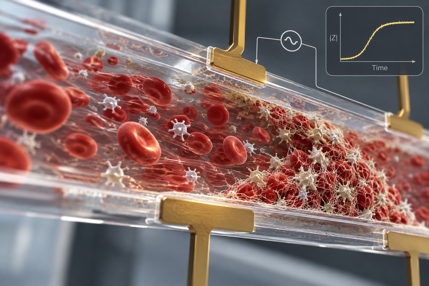
Coagulation under flow
Blood coagulation changes under shear, but most clinical coagulation assays are performed under static or near-static conditions. We develop microfluidic sensors that expose blood to controlled flow while continuously measuring its electrical response.
A major application is extracorporeal support. Patients on ECMO or continuous renal replacement therapy can be at risk of both bleeding and circuit thrombosis, while current measurements provide only intermittent information about coagulation. Our goal is to develop sensors that can monitor these changes closer to the circuit and in real time.
- How does shear rate reshape the electrical signature of clot formation?
- Can emerging thrombotic changes be detected before they produce measurable circuit dysfunction?
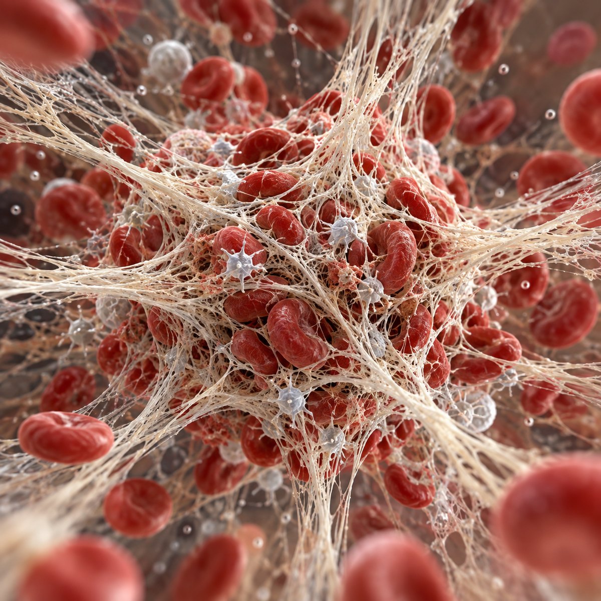
Dielectric sensing of whole blood
The electrical properties of whole blood change as coagulation progresses and cells and fibrin reorganize. We use broadband dielectric measurements to study clot formation, contraction, and structural remodeling beyond conventional clot-time endpoints.
We also study how sensor geometry influences what can be measured. Chamber height, electrode configuration, and sample dimensions can strongly affect sensitivity to contraction. These experiments help distinguish true biological differences from limitations imposed by the sensor itself.
- Which electrical features map onto contractile behavior, and over what range?
- How do inflammatory and uremic environments alter contraction independently of clot time?
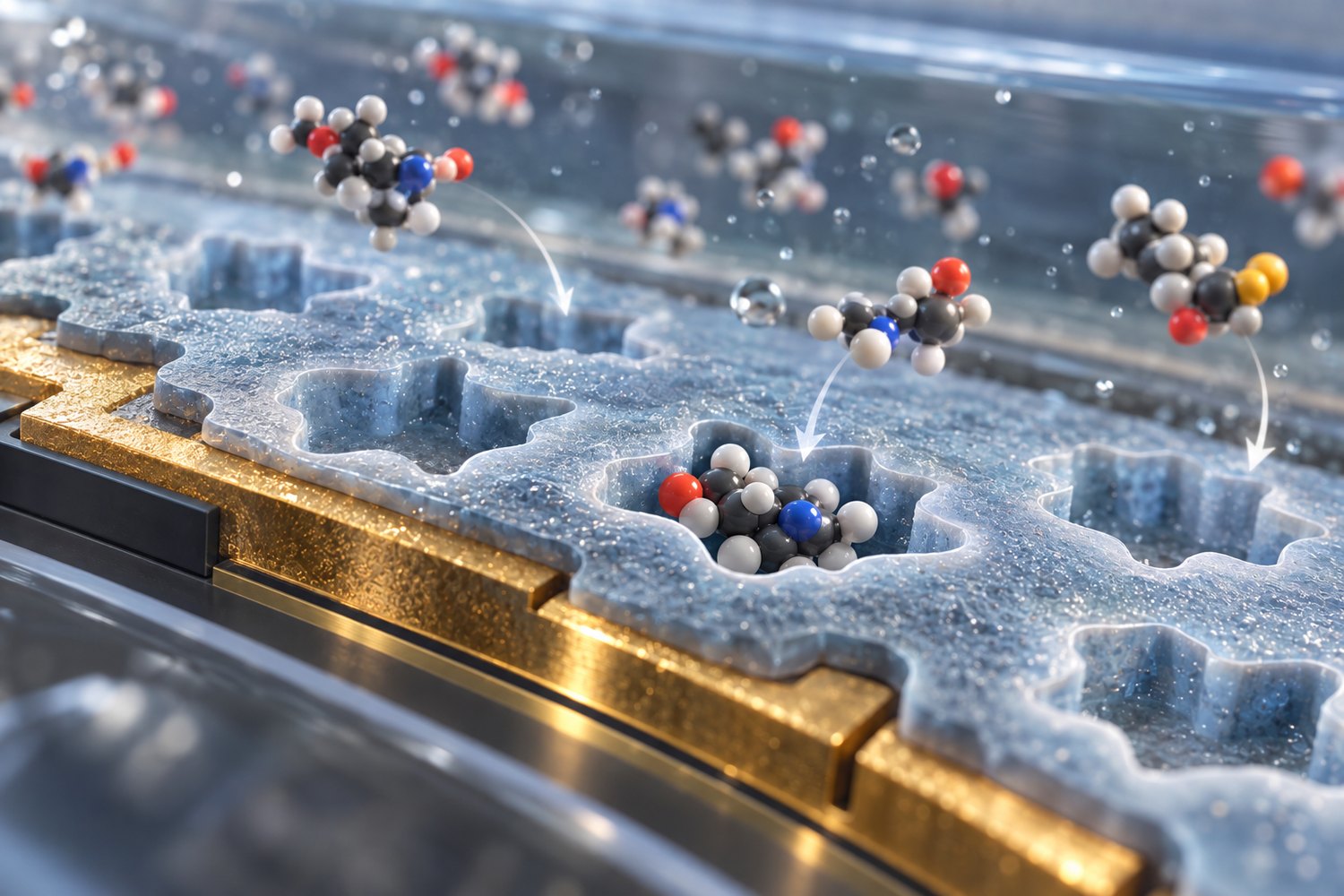
Electrochemical biosensing
We develop nanomaterial-modified and molecularly imprinted electrodes for detecting circulating biomarkers associated with kidney and cardiovascular disease. Current work focuses on protein-bound uremic toxins that accumulate in chronic kidney disease and are poorly removed by conventional dialysis.
We also apply related sensing approaches to other analytical problems, including chemiresistive ammonia detection and impedance-based characterization of milk adulteration. In each case, the sensor interface and measurement strategy are designed around the target and sample.
- How can electrode chemistry provide selective detection of uremic toxins in complex biological samples?
- Can one cartridge report a molecular target and its functional consequence?